Marco TRS 5100 Digital Automated Refraction System
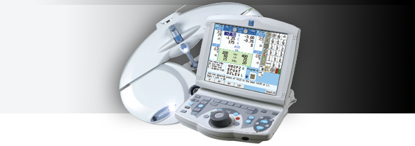
The TRS 5100 benefits from Marco's latest generation of electronic refraction technology. Replacing the standard refractor, it allows practitioners to control the entire refraction process from a keypad small enough to sit in your lap. This keypad also controls the CP-690 Automatic Chart Projector. And because the TRS 5100 is completely programmable, all the lenses are moved for you at the touch of a button, taking you to each new refraction step. While convenient for you, it is also helpful if you delegate refractions and want your technicians to perform the refraction steps in a specific order.
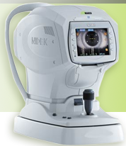
Marco M3 Autorefractor/Keratometer
The New Boston Vision Center, a state of the art Eye Care facility has purchased the newly redesigned M3. The M3 instrument is a three in one devise. The newly redesigned M3 presents several enhanced features such as a smaller footprint, to save even more space, and a new exterior design, but most importantly, the M3 now uses SLD (Super Luminescent Diode) and a highly sensitive CCD device to provide Zonal Ring-Image Technology. This makes it possible to now accurately measure patients with cataracts, corneal opacities, IOLs, and post LASIK.
- Automatic Refraction
- Automatic Keratometry
- Non-Contact Tonometry
- Space Saving
- Time Saving
- High-Speed Measurements
- Accurate Data
- EyeTracking System
- Auto Measure
- Adjustable Monitor
- Motorized Chinrest
- Softer Air Puff
Non-Contact Tonometer, Auto-Refractor and Auto-Keratometer
The Marco M3 non-contact tonometer, auto-refractor, and auto-keratometer combines the measurement of refractive power, corneal curvature and intraocular pressure (IOP) providing fast, highly accurate, reliable measurements with significantly reduced measurement time. Its space saving design eliminates the need for multiple instruments, offering greater efficiency, and it interfaces easily with the patient’s EMR.
Slit Lamp Imaging System (by TelSceen):
Videos and images help patients to understand more, and to understand more quickly. The system is also useful for contact lens fitting (send pictures to the lab for consultation) and for encouraging lens care & replacement compliance – “If you aren’t replacing your lenses on schedule, this picture will show it.” Any patient with a medical condition (keratitis, conjunctivitis, dry eye, etc) will benefit from a visual explanation, and will be more likely to comply with your treatment plan.
Learn more about the TelScreen Slit Lamp Imaging System in this video:
Digital Retinal Imaging & OCT Scans
Our optometrist in New Boston, OH use cutting-edge digital imaging technology to assess your eyes. Many eye diseases, if detected at an early stage, can be treated successfully without total loss of vision. Your retinal Images will be stored electronically. This gives the eye doctor a permanent record of the condition and state of your retina.
This is very important in assisting our eye doctor in New Boston to detect and measure any changes to your retina each time you get your eyes examined, as many eye conditions, such as glaucoma, diabetic retinopathy and macular degeneration are diagnosed by detecting changes over time.
The advantages of digital imaging include:
- Quick, safe, non-invasive and painless
- Provides detailed images of your retina and sub-surface of your eyes
- Provides instant, direct imaging of the form and structure of eye tissue
- Image resolution is extremely high quality
- Uses eye-safe near-infra-red light
- No patient prep required
Digital Retinal Imaging
Digital Retinal Imaging allows your eye doctor to evaluate the health of the back of your eye, the retina. It is critical to confirm the health of the retina, optic nerve and other retinal structures. The digital camera snaps a high-resolution digital picture of your retina. This picture clearly shows the health of your eyes and is used as a baseline to track any changes in your eyes in future eye examinations.
Optical Coherence Tomography (OCT)
An Optical Coherence Tomography scan (commonly referred to as an OCT scan) is the latest advancement in imaging technology. Similar to ultrasound, this diagnostic technique employs light rather than sound waves to achieve higher resolution pictures of the structural layers of the back of the eye.
A scanning laser used to analyze the layers of the retina and optic nerve for any signs of eye disease, similar to an CT scan of the eye. It works using light without radiation, and is essential for early diagnosis of glaucoma, macular degeneration and diabetic retinal disease.
With an OCT scan, doctors are provided with color-coded, cross-sectional images of the retina. These detailed images are revolutionizing early detection and treatment of eye conditions such as wet and dry age-related macular degeneration, glaucoma, retinal detachment and diabetic retinopathy.
An OCT scan is a noninvasive, painless test. It is performed in about 10 minutes right in our office. Feel free to contact our office to inquire about an OCT at your next appointment.
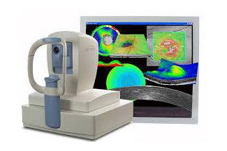
NEW! CIRRUS HD-OCT 4000
 The Cirrus HD-OCT is a non-contact, high resolution tomographic and biomicroscopic imaging device. It is indicated for in-vivo viewing, axial cross-sectional, and three-dimensional imaging and measurement of anterior and posterior ocular structures, including cornea, retina, retinal nerve fiber layer, ganglion cell plus inner plexiform layer, macula, and optic nerve head. The Cirrus normative databases are quantitative tools for the comparison of retinal nerve fiber layer thickness, macular thickness, ganglion cell plus inner plexiform layer thickness, and optic nerve head measurements to a database of normal subjects. The Cirrus HD-OCT is intended for use as a diagnostic device to aid in the detection and management of ocular diseases including, but not limited to, macular holes, cystoid macular edema, diabetic retinopathy, age-related macular degeneration, and glaucoma.
"Being able to visualize so quickly areas of macular ischemia in my DR patients without the use of a contrast agent could really help me more easily select patients with DME eligible for treatment." - Prof. JF Korobelnik, University Hospital Pellegrin, Bordeaux, France
Visualization at the speed of CIRRUS
Analyzing a single pathology from multiple views provides comprehensive insight and analysis of the clinical situation. How this helps you:
- Spot small areas of pathology. Tightly spaced B-scans, (either 30 or 47 μm apart), in the cube ensure that small areas of pathology are imaged. For reference, a human hair is about 40-120 μm in diameter.
- Visualize the fovea. Scans that are spaced further apart than in the CIRRUS cube may miss the central fovea.
- Fuel for analysis. Millions of data points from the cube are fed into the Zeiss proprietary algorithms for accurate segmentation, reproducible measurements and registration for change analysis.
- Take the pressure off the operator. As long as the scan is placed in the vicinity of the fovea or optic nerve, the software automatically centers the measurements after the capture.
- See the tissue from different perspectives. View the cube data from all angles, with 3D rendering, OCT fundus images and Advanced Visualization™.
- Future ready. Previously captured CIRRUS cubes can be analyzed using new analyses.
Visual Field Testing
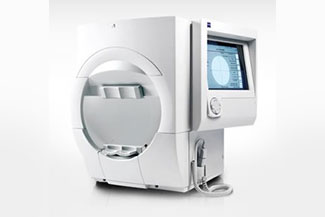
A visual field test measures how much 'side' vision you have. It is a straightforward test, painless, and does not involve eye drops. Essentially lights are flashed on, and you have to press a button whenever you see the light. Your head is kept still and you have to place your chin on a chin rest. The lights are bright or dim at different stages of the test. Some of the flashes are purely to check you are concentrating.
Each eye is tested separately and the entire test takes 15-45 minutes. Your optometrist may ask only for a driving licence visual field test, which takes 5-10 minutes. If you have just asked for a driving test or the clinic doctor advised you have one, you will be informed of the result by the clinic doctor, in writing, in a few weeks.
Normally the test is carried out by a computerized machine, called a Humphrey. Occasionally the manual test has to be used, a Goldman. For each test you have to look at a central point then press a buzzer each time you see the light.

